Index
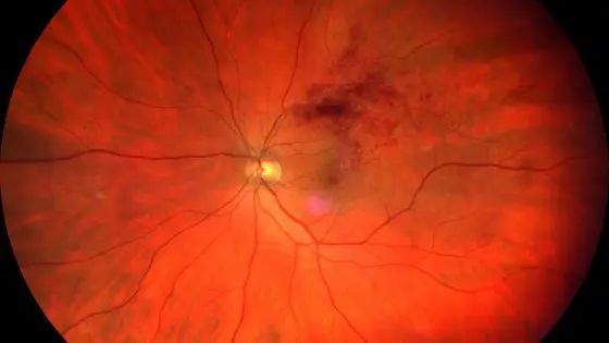
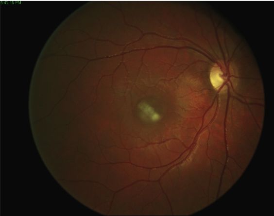
Central retinal arterial occlusion is caused by an embolus or prolonged spasm of the retinal artery walls. This spasm causes a blockage of blood flow in the central retinal artery.
The embolus that causes retinal arterial occlusion can originate at the level of the carotids, in the presence of arteriosclerosis (which represents one of the main risk factors for this ocular pathology), but also at the heart level in the presence of certain pathologies.
Central retinal arterial occlusion occurs more frequently, but not exclusively, in older people, in people of the male gender, and in people with clotting factor mutations. The consequences of central retinal artery occlusion are serious and irreversible. Precisely for this reason it is necessary to pay particular attention to prevention through theidentification and control of risk factors associated with it.
Risk factors
Risk factors most associated with central retinal artery occlusion include: arterial hypertension; platelet disorders; carotid artery disease; hypercholesterolemia; atherosclerosis; giant cell arteritis; diabetes; hereditary haematological factors; the use of oral contraceptives or a pre-existing event of central retinal artery occlusion in one eye. In addition, people who have one or more risk factors for retinal arterial occlusion must develop, with their ophthalmologist and general practitioner, a prevention protocol that includes the adoption of an adequate lifestyle and any pharmacological treatments. aimed at lowering the risk.
Treatment
There are now several first aid therapeutic approaches for central arterial occlusion which primarily include the digital massage of the eyeball with closed eyelid, trying to dislocate the embolus in a branch of the artery or even in one of its capillaries in such a way as to reduce the area of ischemic retinal damage.
In addition, in recent years one is experimenting with a fibrinolytic therapy, aimed at eliminating the embolus, e vasodilator drugs o cortisone. Unfortunately, however, these therapies are inefficient in the vast majority of cases and in any case must be implemented in the first 4-6 hours after the onset of symptoms.
Pathology and treatment on video
Do you need more information?
Do not hesitate to contact me for any doubt or clarification. I will evaluate your problem and it will be my concern and that of my staff to answer you as quickly as possible.

