Index
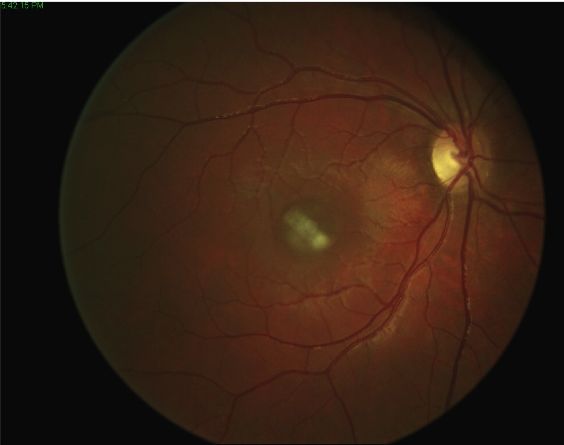
Il macular hole is a dangerous eye disease that consists in the loss of retinal tissue at the level of the macula, the most central part of the retina responsible for clear and detailed vision.
Why and how it originates
Macular hole is usually caused by a traction force persistently exerted on the macula. There are conditions that can favor the formation of a macular hole, among these we find vitreomacular traction (VMT), myopia magna, la proliferative diabetic retinopathy (PDR), diabetic macular edema, post-operative macular edema and persistent intraocular inflammation. The traction force exerted on the macula may initially result in a circumscribed elevation of the sensorineural retina, ie the retinal layer containing the photoreceptors (cones and rods). In some cases the traction force resolves spontaneously, in other cases, however, the traction persists and / or increases, causing a tear in the central part of the retinal tissue and the formation of the macular hole.
Symptoms
When the traction on the macula causes it to lift, the patient feels one blurred and distorted central vision. If the traction persists over time or increases in intensity, the risk of a macular hole formation becomes high. If no action is taken, a tear can be created, a real break in the retinal tissue, which is visually perceived as a blind spot (scotoma) in central vision.
The formation of a macular hole represents a real eye emergency, since if you do not intervene immediately, the visual damage can become permanent and turn into a irreversible loss of central vision. In addition, other very serious complications can be generated, such as secondary retinal detachment.
The diagnosis
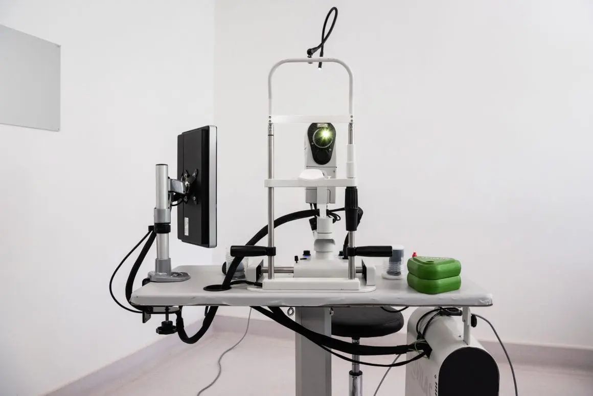
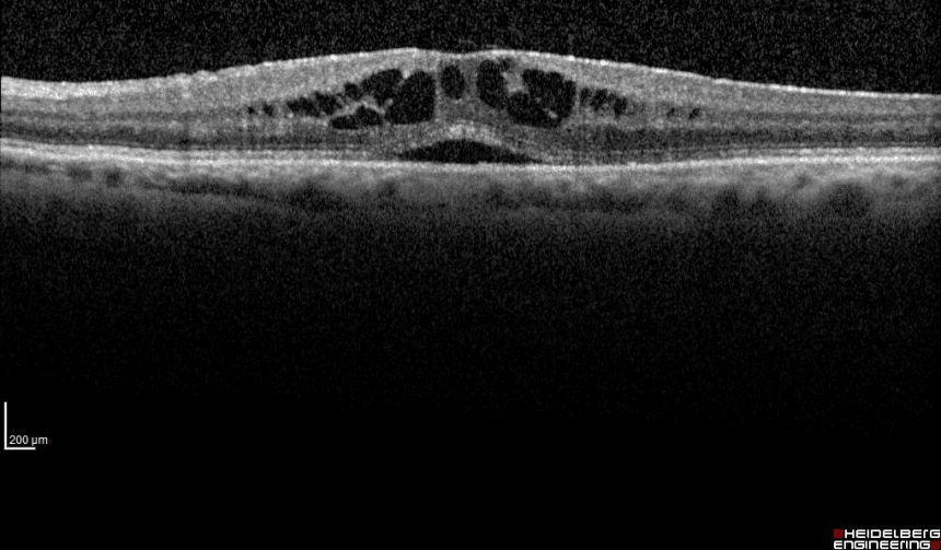
Macular hole can be diagnosed on slit lamp ophthalmoscopy thanks to the Watzke-Allen test. The optical coherence tomography (OCT) high resolution it also allows to analyze the complex structure of the retina at the level of each single cellular layer that composes it. The computerized field of view , microperimetry allow to accurately evaluate the functionality of the retina in the central (macular) part.
In the presence of a vitreous body that is not perfectly transparent, for example in the case of hemovitreus, it is necessary to perform aB-Scan ultrasound to verify the possible existence of a vitreous traction on the retina due to natural causes or trauma. Vitreous traction must be monitored very carefully to rule out not only macular hole formation but also other serious complications such as retinal detachment.
The treatment
Once diagnosed and quantitatively evaluated thanks to OCT, the macular hole must be treated promptly by means of an intervention of vitrectomy via pars plana. This surgery involves the removal of the vitreous body and epiretinal membranes, including the internal limiting membrane of the retina, and is essential to restore central vision or at least safeguard residual vision and avoid the risk of retinal detachment.
Il time between the formation of the macular hole and the operation it must be as short as possible, since when a macular hole is formed the photoreceptors of the macula lose contact with the cells of the retinal pigment epithelium, a source of oxygen and nutrients, and quickly undergo cell death, making the loss of central vision irreversible .
After the vitrectomy, the eye gradually recovers the lost vision and theextent of visual recovery depends, as well as on timeliness with which it was possible to intervene, from size of the macular hole and by the patient's ability to maintain a position that facilitates healing of the macula, defined position face down.
Location Face Down
At the end of the vitrectomy, a gaseous tamponade medium able to exert a pressure force that favors the repositioning of the macula and its re-attachment to the back of the eye, thus obtaining closing the hole. In order for the gas bubble to press correctly on the macula it is necessary for the patient to keep his head with his face parallel to the floor, the so-called "position face down".
Correct maintenance of the position face down in the post-operative period it is as fundamental for the successful outcome of the treatment of the macular hole as the success of the surgery itself.
Assorurale's position face down must be kept for at least 3 4-days for the as many hours a day as possible. At the end of the convalescence, the eye recovers the lost vision in a progressive way and the extent of the recovery can be evaluated in the follow-up.
If you want to know the most comfortable and effective positions to take during your convalescence, we suggest you download the following Ebook.
The macular hole and the importance of position face down: a guide for patients undergoing vitrectomy

Pathology and treatment on video
Do you need more information?
Do not hesitate to contact me for any doubt or clarification. I will evaluate your problem and it will be my concern and that of my staff to answer you as quickly as possible.


