Index
Closed-angle glaucoma is a pathology caused by high intraocular pressure which can cause irreversible damage to the optic nerve.
This disease is caused by a pathological increase in the intraocular pressure (Iinoasshole Pcracks, IOP), due to an increase in production in the amount of aqueous humor present in front room eye or a difficulty in its outflow.
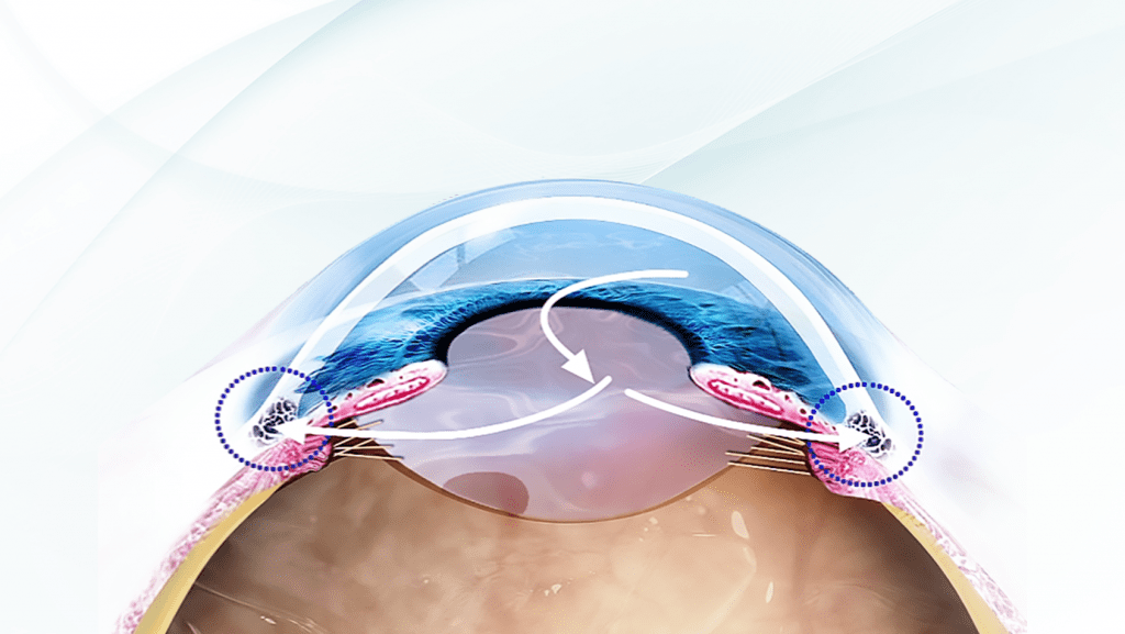
The aqueous humor is a transparent liquid that has the function of nourishing the cornea and crystalline and to eliminate waste products. In a healthy eye, it is continuously produced within the anterior chamber of the eye, by the cells of the non-pigmented epithelium of the ciliary body. Aqueous humor is constantly drained - at the same rate at which it is produced - through an ocular structure called the trabeculum scleral horny and other anatomical structures assigned to it.
Thanks to the correct functioning of this physiological mechanism, in a healthy eye the IOP maintains rather constant values ranging between 14 and 21 mmHg
Il closed-angle glaucoma arises when the drainage mechanism of the aqueous humor is progressively blocked. In some cases, the iris is pushed forward enough to occlude the drainage angle and completely blocks the outflow of aqueous humor. This block determines a very sudden increase in IOP and of such magnitude as to cause an attack of acute glaucoma, which represents a real ophthalmic emergency.
Symptoms
Symptoms of an acute glaucoma attack can consist of the presence of disorders visual (sudden and severe lowering of vision, seeing colored halos around lights) accompanied by redness and severe eye pain, nausea and vomiting.
An acute glaucoma attack is a dangerous event for vision, as it can lead to complete and irreversible blindness in a very short time, if not adequately and promptly treated.
Risk factors
There are generally no warning symptoms of an acute glaucoma attack, so it is very important to consider the existence of factors risk for this serious illness. Among the most important risk factors we find inheritance,old age, hyperopia high, and the presence of a cataract especially if associated with an intumescent lens.
In people at risk, symptoms such as blurry vision and seeing halos around lights accompanied by occasional headaches and slight pain or redness in the eye could be considered warning signs of an acute glaucoma attack. In these cases it is necessary to be examined urgently by an ophthalmologist.
Diagnosis
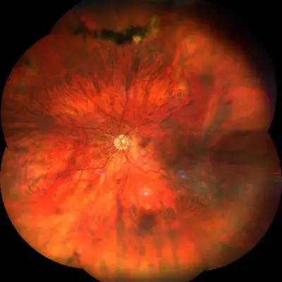
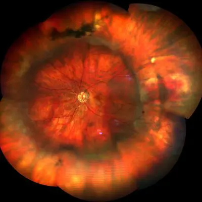
Closed-angle glaucoma, if an acute attack has not occurred, progresses slowly and quietly. Often the patient does not present specific symptoms except in the most advanced stages of the disease with a lowering of central vision and a reduction of the peripheral visual field.
So you have to focus on the prevention that has to be done through the monitoring of pressure intraocular (IOP) and the maintenance of its values within physiological limits thanks to the daily use of special eye drops.
Patients at risk should be monitored regularly with a complete eye examination and instrumental examinations such as Perimetry (computerized or manual) which serves to analyze the total retinal function therefore both central and peripheral and the Tomography a Coherence Optical (OCT) ad high resolution, which allows you to examine the optic nerve and the retinal nerve fiber layer (RNFL, from English Rethinal Nherb FIberian Layer).
If glaucoma develops and the optic nerve shows pain, urgent action is needed. In patients with an anatomical or familial predisposition for an acute glaucoma attack, anatomical features should be carefully measured angle corneoscleral. It is also necessary to verify the thickness of the lens by means of a diagnostic A-scan ultrasound to avoid that its dangerous intumescence cannot be adequately considered.
Treatment
The therapy to be practiced urgently is iridotomy laser. This surgery para-surgical is employed in the patient with a acute glaucoma attack and with closed-angle glaucoma with an evolutionary trend.
These conditions are caused by a irido-lenticular block followed by a sudden and abrupt raising of the intraocular pressure (IOP). Such conditions are extremely dangerous as they can lead to complete and irreversible blindness in a very short time.
Furthermore, this type of treatment is also indicated in those patients who have predisposing anatomical risk factors (such as a lower room).
Laser iridotomy
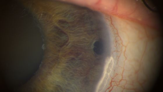
The laser iridotomy surgery involves the creation of a small hole at the level of the iris that acts as a via drainage for the aqueous humor. This hole, located in the anterior chamber of the eye, allows the balance between the incoming and aqueous humor to be restored aqueous humor outgoing normalizing the intraocular pressure.
Laser iridotomy is performed on an outpatient basis, generally in a suitably equipped eye clinic. The eye to be treated is anesthetized by applying eye drops and then a lens is applied that allows the area to be treated to be focused.
The hole created in the iris is the size of a pinhead and in most cases it is not visible because it remains hidden under the upper eyelid. The procedure takes a few minutes and is not invasive. Due to the blurry vision that persists for a few hours, the patient is advised to be accompanied by a person who can take him home after the surgery.
Pathology and treatment on video
Do you need more information?
Do not hesitate to contact me for any doubt or clarification. I will evaluate your problem and it will be my concern and that of my staff to answer you as quickly as possible.


